Norwich Image Interpretation Course

Heidi Nunn (Advanced Practice Reporting Radiographer)


The Hand

Phalanges Metacarpals Thumb Paediatric fractures Enchondroma

(hover over images to zoom, click to enlarge)

Learning outcomes
  • Assess skeletal radiographs using a systematic approach
  • Understand the importance of obtaining the correct radiographic projections to demonstrate the thumb, metacarpals and phalanges
  • Describe metacarpal and phalangeal anatomy
  • Understand mechanisms of injury and the likely fractures/dislocations which may result
  • Search for subtle injuries and understand their clinical significance
  • Accurately describe dislocations and associated fractures
  • Understand common eponyms
  • Know the common avulsion sites and ligament injuries
  • Understand the paediatric Salter-Harris classification
  • Recognise and understand the relevance of common lytic lesions
Phalanges
  • Small avulsion fractures are common, easily missed and are functionally important. Avulsions may be seen:
    1. On the DP view - at the head or base of the phalanges and head of the adjacent metacarpal. This is at the insertion of the medial and lateral collateral ligaments, and the resultant avulsed fragment may be very subtle:
Avulsion fracture at insertion of collateral ligament
    1. On the lateral view: anterior aspect - at the head or base of the phalanges or adjacent metacarpal (most commonly at the base of the middle phalanx). This is at the insertion of the volar plate (which refers to the joint capsule). This injury is caused by hyperextension:
Avulsion fracture at insertion of volar plate
    1. Again on the lateral, but on the dorsal aspect, at the insertion of the extensor tendon:
Avulsion fracture at insertion of extensor tendon
    1. This tendon may rupture without an attached bone fragment and the resulting flexion deformity at the distal interphalangeal joint is called a mallet finger:
Mallet deformity
  • It is important to obtain a lateral view of the affected finger when injury to the phalanges is suspected, as an oblique does not demonstrate small avulsions or the extent of displacement of larger avulsion fractures:
Volar plate avulsion fracture of index finger ... no lateral

  • Dislocations to the interphalangeal joints are common. The dislocation is described by which joint is affected and by the direction of the phalanx distal to the joint relative to the phalanx proximal to the dislocated joint (eg, dorsal dislocation at the PIPJ). It is important to assess for any associated fractures pre- and post-reduction:
Dorsal dislocation PIPJ with avulsed fragment
Metacarpals
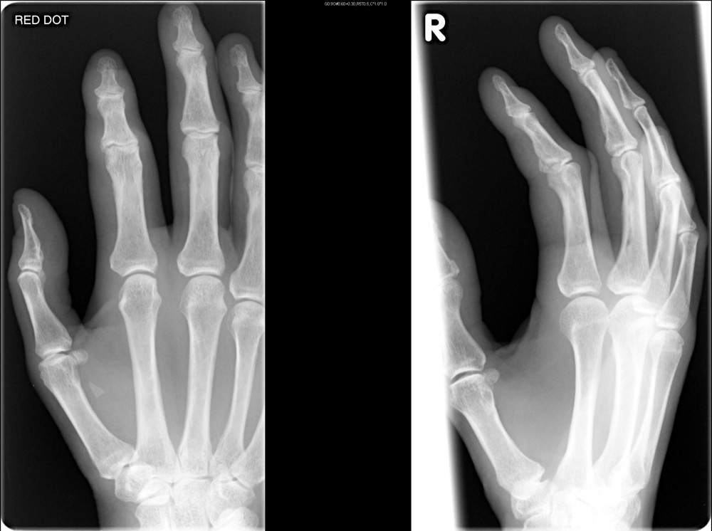
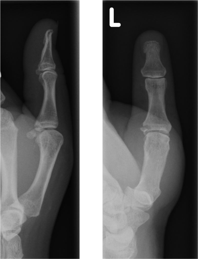
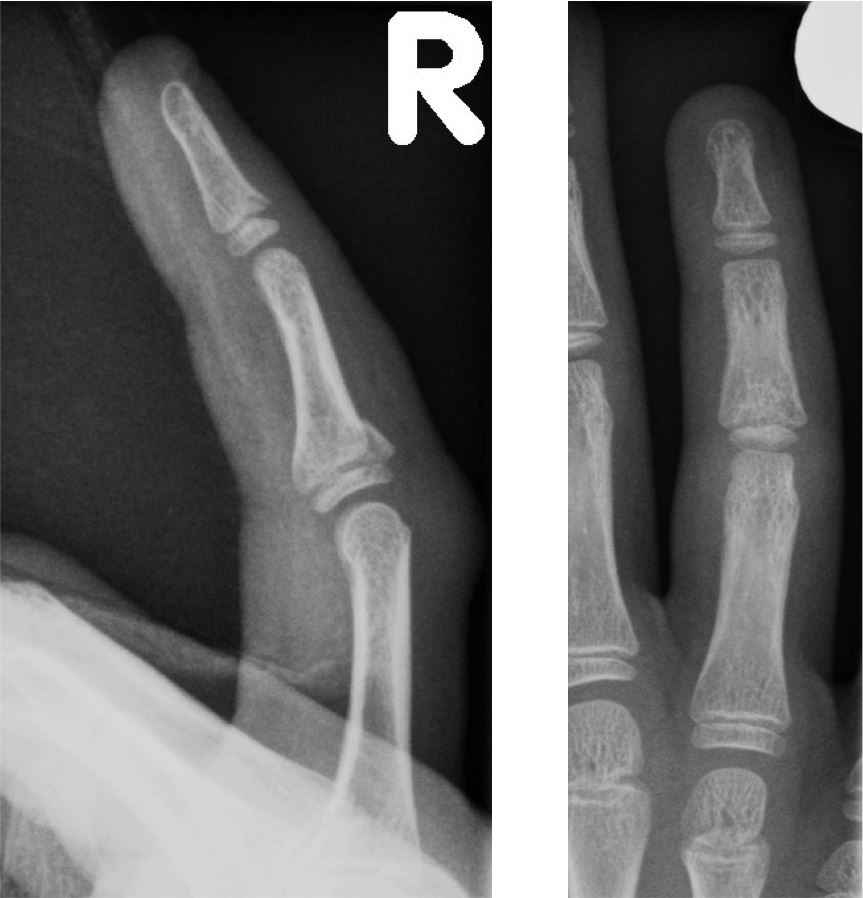
  • Fractures to the shaft, neck or head, particularly of the 4th or 5th metacarpal are common. Most often caused by punching injuries. and are rarely missed as they are usually displaced:
Fracture distal shaft 5th metacarpal
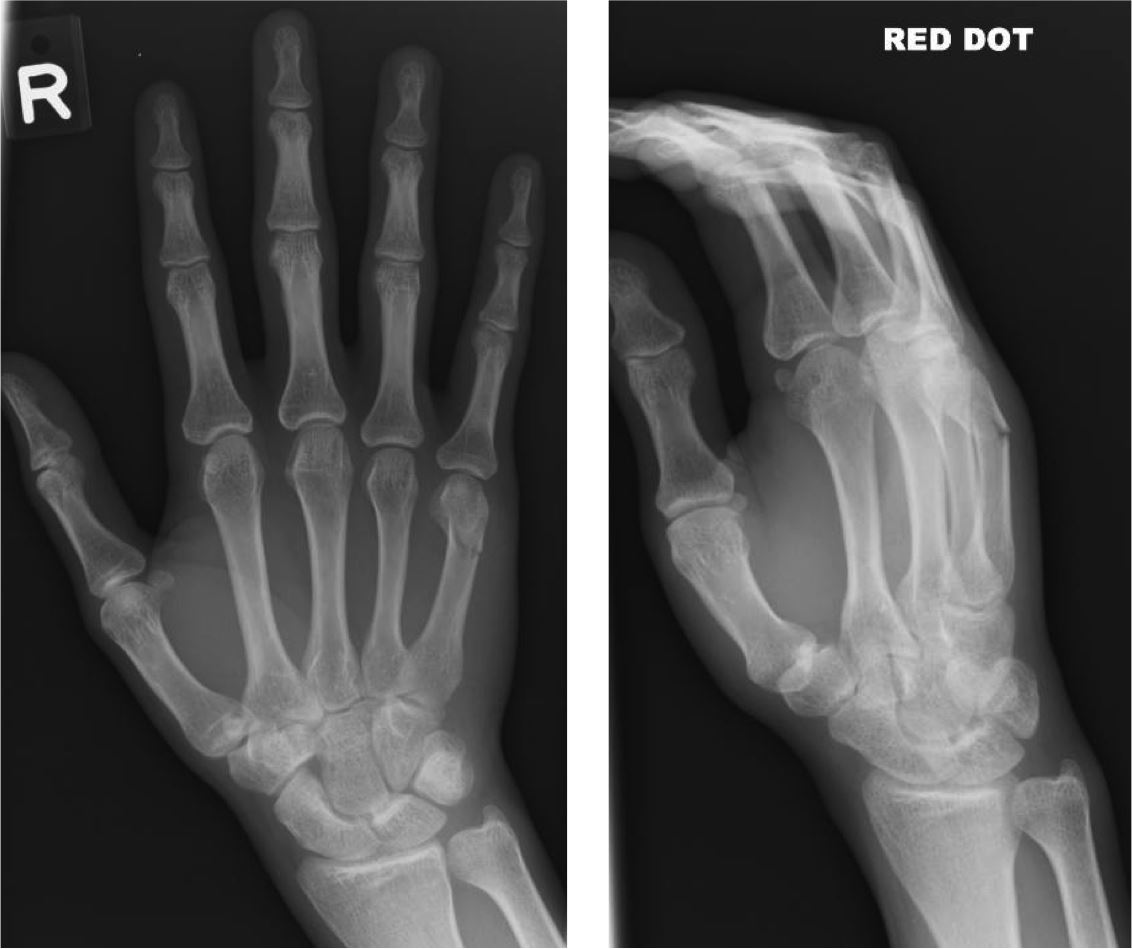
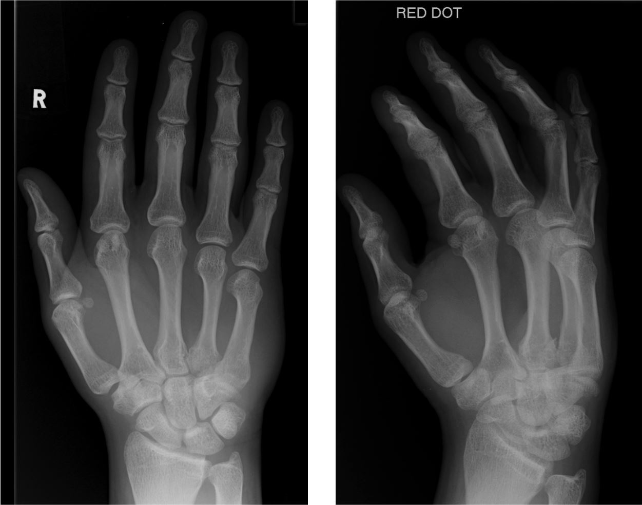
  • More subtle, and yet clinically significant, are fractures to the base of the 4th or 5th metacarpal:
Fracture base 5th metacarpal
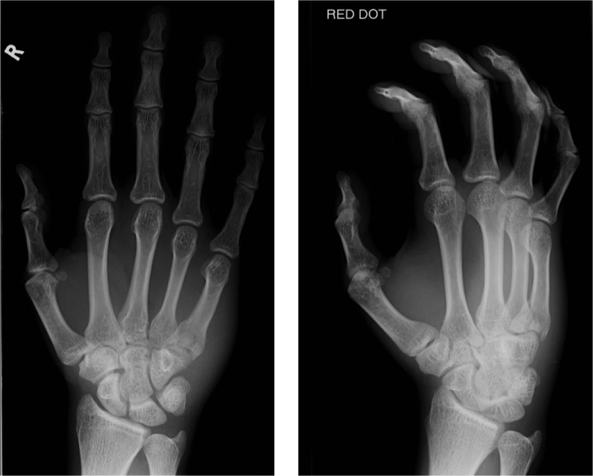
  • These are often associated with dislocation to the 4th or 5th carpo-metacarpal joints. A dislocation can be identified by recognising loss of the normal joint space (2mm) at the metacarpal base:
Fractured 4th metacarpal, dislocation 5th carpo-metacarpal joint
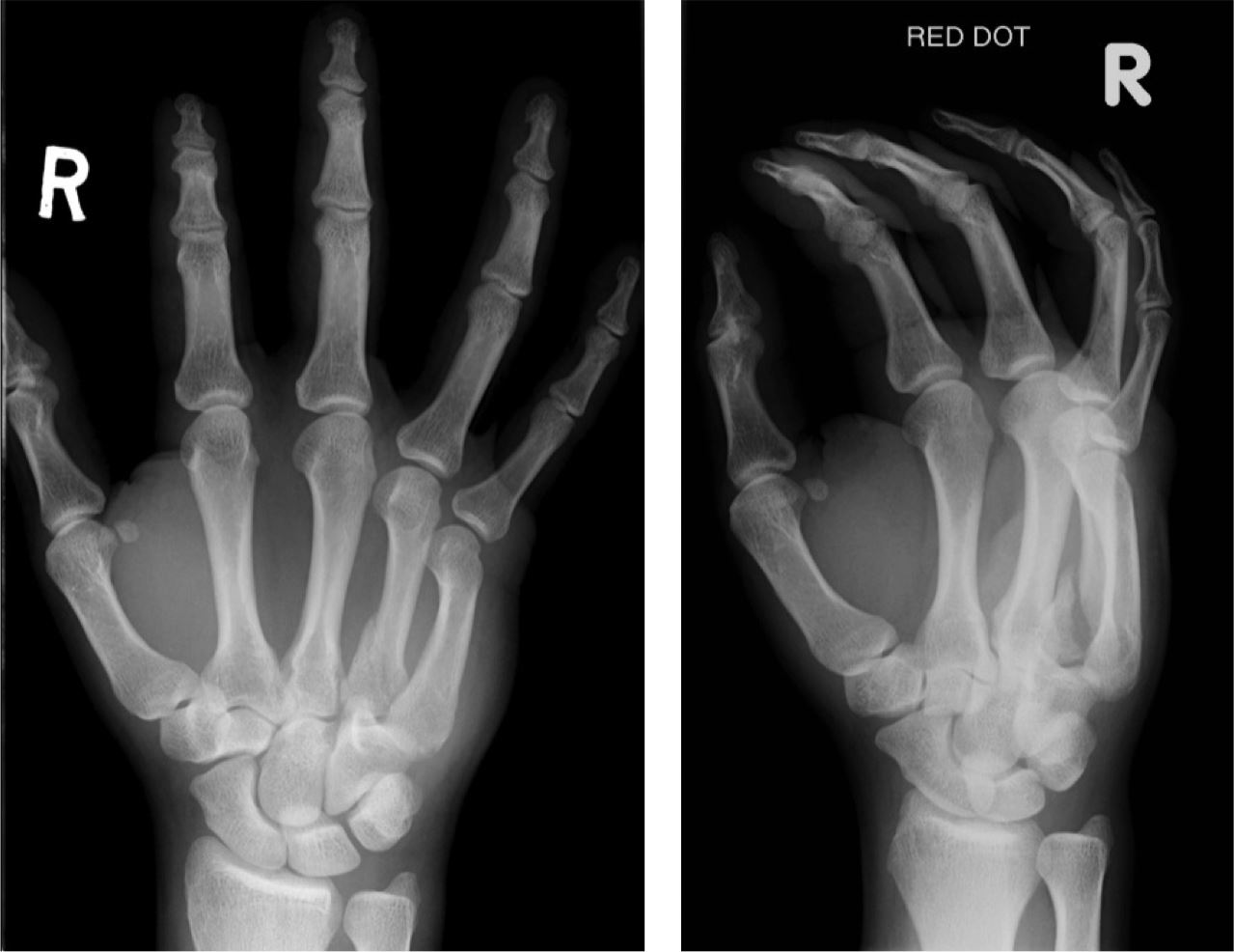
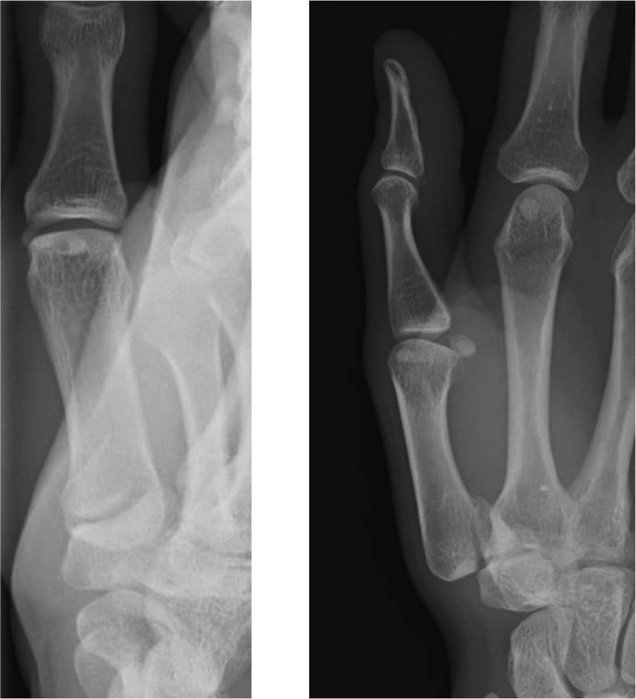
  • Another associated injury is to the hamate, often evident on the oblique image:
Fracture-dislocation 4th/5th plus fractured hamate
Mechanism of injury is similar to the 5th metacarpal neck fracture so, when index of suspicion is high and a fracture to the head or neck isn't present, scrutinise the base of the 4th and 5th metacarpals, and the adjacent hamate.
Thumb
  • A common fracture at the base of the 1st metacarpal is an oblique intra-articular fracture, usually with dorsal subluxation of the shaft. This is the Bennett's fracture-dislocation:
Bennett's fracture-dislocation
  • Similar to the Bennett's fracture-dislocation, but comminuted, is the Rolando's fracture-dislocation. Both are unstable injuries:
Rolando's fracture-dislocation
  • Ligament injuries are also common around the thumb, especially at the insertion of the ulnar collateral ligament. This is at the medial aspect of the base of the proximal phalanx. This is termed "skier's thumb":
Avulsion fracture at insertion of ulnar collateral ligament
Paediatric Fractures
  • Salter-Harris fractures involving the physeal growth plate and adjacent metaphysis and/or epiphysis are common. An injury to this area will cause a fracture to the weakest point, which is the cartilaginous growth plate, and spare the stronger joint capsule, ligaments and tendons. Depending on the involvement of the adjacent metaphysis and epiphysis, determines the classification:
    • Salter-Harris Type I = Injury through the physeal growth plate only, usually with displacement. No involvement of the adjacent metaphysis or epiphysis.
    • Salter-Harris Type II = Fracture extending through the physeal growth plate and adjacent metaphysis:
Salter-Harris II
    • Salter-Harris Type III = Fracture line running through the growth plate and adjacent epiphysis, which extends into the joint.
    • Salter-Harris Type IV = Fracture involving the epiphysis, physeal plate and metaphysis.
    • Salter-Harris Type V = A compression fracture extending through the physeal growth plate. Often difficult to detect.

    As the classification increases from I to V, the frequency of injury decreases (types I and II common) but the severity of injury increases.

  • Subtle torus fractures are also common, particularly at the base of the phalanges. Torus fractures refer to buckling of the cortex, with little displacement:
Torus fractures
Enchondroma
  • A benign lytic lesion, often seen within the phalanges. Usually asymptomatic and an incidental finding, but can fracture. Characteristically lobulated, slightly expansile with endosteal scalloping and cortical thinning. Multiple enchondromas also commonly seen (Ollier's disease).
Enchondroma with pathological fracture

TAKE THE HAND SELF-TEST

- return to top -