Norwich Image Interpretation Course

Heidi Nunn (Advanced Practice Reporting Radiographer)


The Knee

Ottawa rules Suprapatellar bursa Patella Tibial plateau Segond fracture Intercondylar eminence Proximal fibula Osteochondritis dissecans Osgood-Schlatter's disease Pellegrini Stieda Synovial osteochondromatosis Diaphyseal aclasis Non-ossifying fibroma

(hover over images to zoom, click to enlarge)

Learning outcomes
  • Understand Ottowa rules for knee imaging
  • Assess skeletal radiographs using a systematic approach
  • Describe knee anatomy
  • Recognise and understand the significance a suprapatellar effusion and lipohaemarthrosis
  • Understand mechanisms of injury and the likely fractures/dislocations which may result
  • Search for subtle injuries and understand their clinical significance
  • Accurately describe dislocations and associated fractures
  • Understand common eponyms
  • Recognise potential ligament injuries
  • Understand paediatric anatomy
  • Recognise common pathological conditions seen around the knee
Ottawa rules
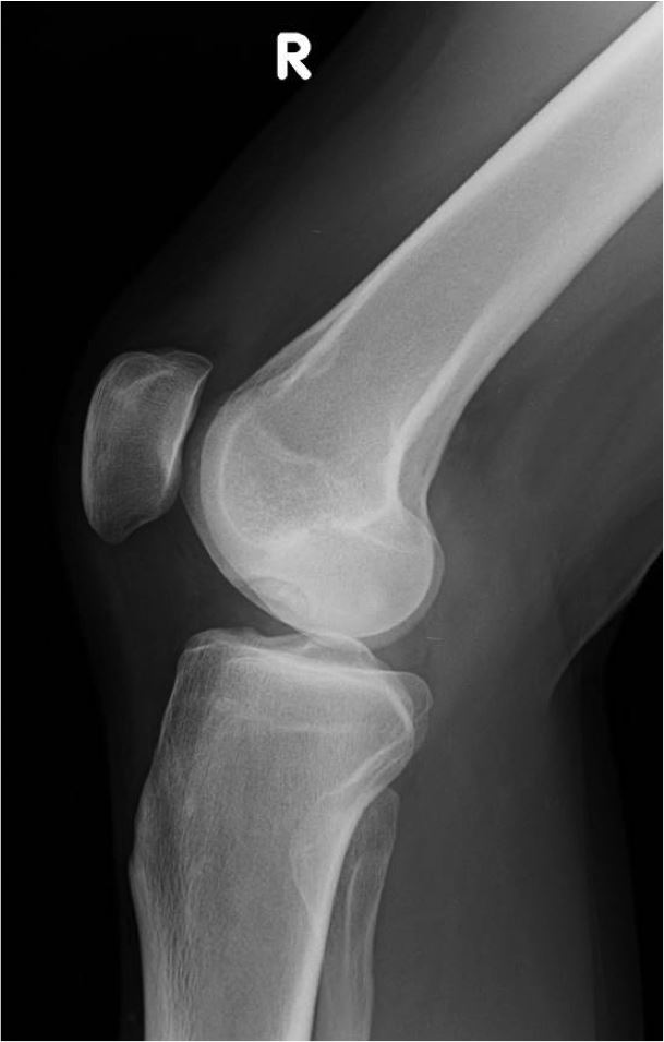
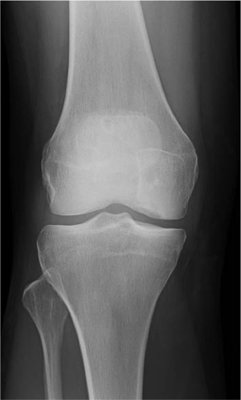
State that a knee radiograph is only required for patients with knee injuries with any of the following:
  • Age 55 or over.
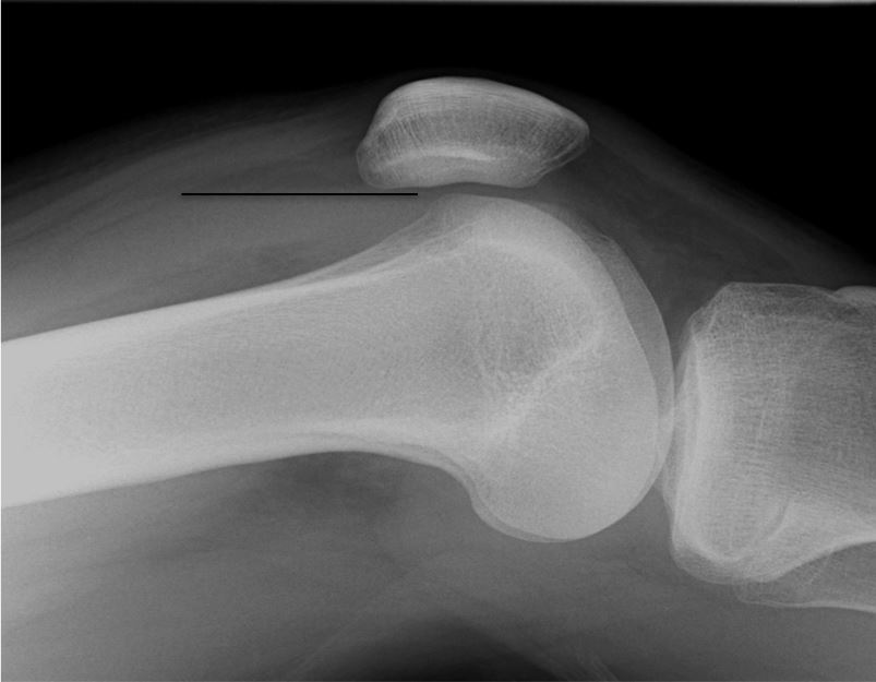
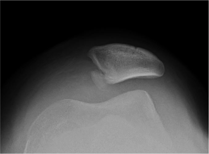
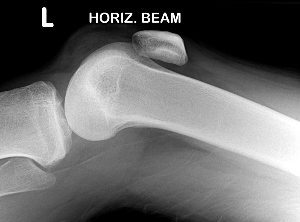
  • Isolated tenderness of the patella (no bone tenderness of the knee other than the patella).
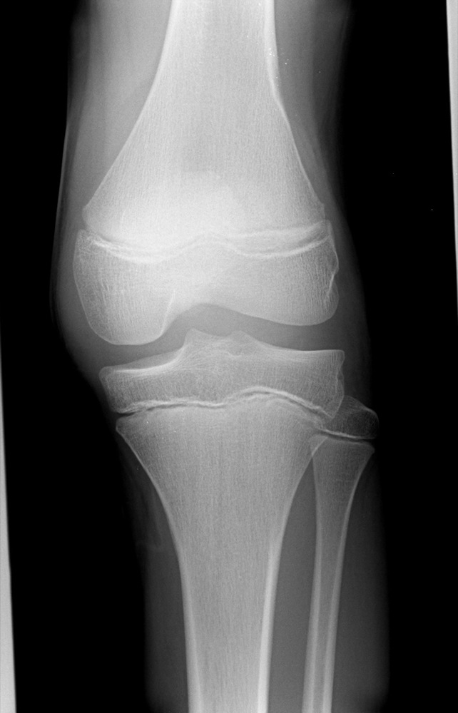
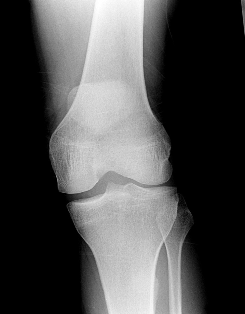
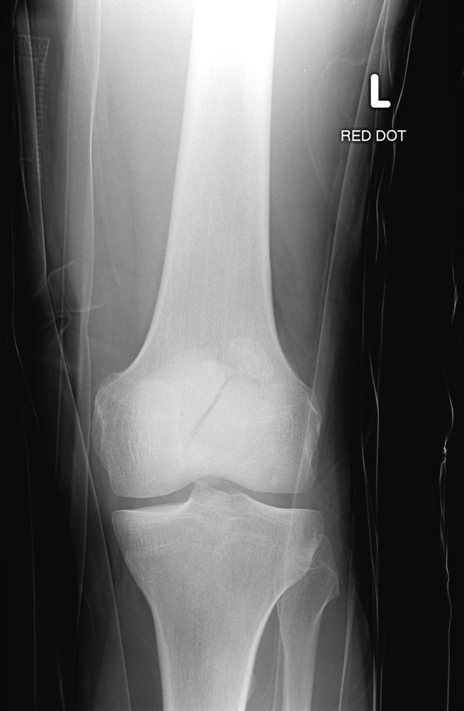
  • Tenderness at the head of the fibula.
  • Inability to flex to 90 degrees.
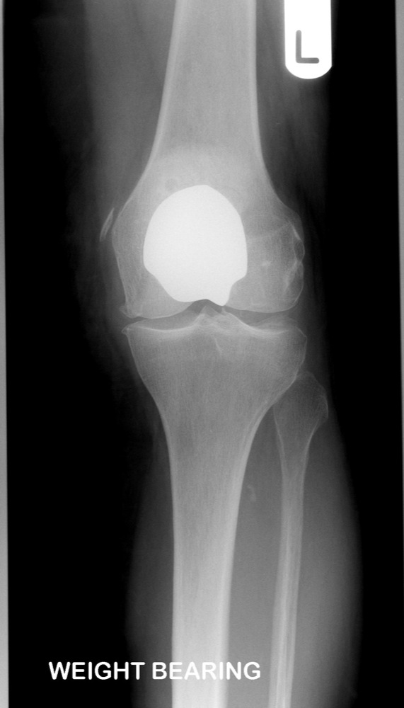
  • Inability to weight bear both immediately and in the casualty department (ie, 4 steps - unable to transfer weight twice onto each lower limb regardless of limping).
Suprapatellar Bursa
  • The fat pad posterior to the quadriceps tendon is divided into anterior and posterior compartments by a soft tissue density; the suprapatellar bursa. This should measure 5mm or less:
Normal bursa   Normal bursa
  • This is located within the joint capsule, therefore it may expand to form a joint effusion (>5mm). This is indicative of soft tissue injury (meniscal or ligamentous). The majority of knee injuries affect the soft tissues, with no bony injury demonstrated:
Effusion, no fracture   Effusion, no fracture
  • Intra-articular fractures will cause leakage of blood and marrow into the joint which will again distend the bursa to form a lipohaemarthrosis. Fat (lucent) lies on top of the fluid (blood), which is radiographically dense. Indicates a fracture even if one is not seen.
Lipohaemarthrosis, no fracture seen on this image   Lipohaemarthrosis, no fracture seen on this image
Patella
  • Fractures may be comminuted, transverse or vertical. Sometimes a skyline projection may be required to demonstrate a vertical fracture, however, this is contraindicated for transverse fractures, as flexing the knee will cause the fracture to displace:

  • undisplaced:
Undisplaced fracture patella   Undisplaced fracture patella, plus lipohaemarthosis

  • displaced:
Displaced fracture patella   Displaced fracture patella
  • Skyline views can also be useful to demonstrate subtle subluxations:

    Lateral subluxation patella   Lateral subluxation patella plus lipohaemarthrosis   Lateral subluxation patella
  • Look for osteochondral fractures associated with patella dislocations, ie, medial patella or lateral femoral condyle:

    Avulsion fracture following dislocated patella    Avulsion fracture medial patella and lateral subluxation of patella
  • Ruptures of the infrapatellar ligament may occur. Evidence of this is an increased distance between the inferior pole of the patella and the tibial tuberosity. This distance normally should be roughly the length of the patella, plus or minus 20%.

    Rupture infrapatellar ligament   Rupture infrapatellar ligament
  • Bipartite patella is often seen, which is an unfused secondary ossification centre. This is characteristically located at the supero-lateral aspect and should not be mistaken for a fracture. However, a bipartite patella may also fracture:

    Bipartite patella plus fractured patella    Bipartite patella plus fractured patella
Tibial Plateau
  • Fractures are associated with ruptures of the medial collateral ligament and anterior and posterior cruciate ligaments.
  • Lateral tibial plateau is the most common site. Can be subtle and may be easily overlooked. However, this is a significant injury:
Lateral tibial plateau fracture   Lateral tibial plateau fracture plus liphaemarthrosis
  • An imaginary line drawn along the lateral femoral condyle and the medial cortex of the proximal fibular shaft should not have more than 5mm of tibial condyle located laterally. If more than this is seen laterally, a split/depression fracture should be suspected:
Normal knee    Fracture lateral tibial plateau
  • Look closely for the lucent fracture line (split) and an area of increased bone density / sclerosis indicating impaction (depression):
Lateral tibial plateau fracture   Lateral tibial plateau fracture
  • Because the tibial plateau slopes posteriorly 15 degrees, it can be difficult to determine the degree of depression. Displacement is therefore often underestimated.
Segond fracture
  • Refers to a characteristic linear fragment arising from the lateral margin of the lateral tibial plateau. This fragment is attached to the lateral capsular ligament and is associated with disruption of the anterior cruciate ligament. It is a more significant injury than it initially appears:
Segond fracture    Segond fracture and fracture head of fibula    Segond fracture
Intercondylar Eminence (Tibial Spine)
  • Avulsion fractures occur at the insertion of the anterior cruciate ligament (most common) and posterior cruciate ligament. The avulsion may be a partial avulsion or complete detachment. Most commonly seen in young adults:
ACL avulsion fracture
PCL avulsion fracture
Proximal Fibula
  • Fibular head and neck fractures are usually accompanied by lateral tibial plateau fractures or knee ligament injuries (cruciate or collateral ligaments). As with the segond fracture, an apparently small fracture in this region is often more significant than it appears:
Fracture fibular head    Fracture fibular head plus liphaemarthrosis
  • Fractures seen more distally at the proximal fibular shaft are often accompanied by ankle injuries (Maisonneuve):
Maisonneuve fracture    Maisonneuve fracture
Osteochondritis Dissecans
  • Refers to a subarticular lucency with sclerotic margins, within which is seen an oval segment of bone (area of avascular necrosis). This will often give symptoms similar to the presence of a loose body, as there subsequently becomes a gradual separation of this loose bone:
Osteochondritis dissecans    Osteochondritis dissecans
  • Osteochondritis dissecans is characteristically seen at the lateral aspect of the medial femoral condyle. Other common sites may be within the talus, or the capitellum:
Osteochondritis dissecans - talus    Osteochondritis dissecans - capitellum
Osgood-Schlatter's Disease
  • Osteochondritis of the tibial tuberosity. Occurs in adolescents with repeated trauma to the knee. The normal ossification centre for the tibial tuberosity may demonstrate a wide variety of normal variants and is often fragmented. Diagnosis is made clinically, and radiography is not indicated.
Pellegrini-Stieda
  • Refers to post-traumatic calcification within the medial collateral ligament. Should not be mistaken for an acute injury:
Pellegrini-Stieda    Pellegrini-Stieda
Synovial Osteochondromatosis
  • Refers to multiple cartilaginous loose bodies, which calcify within the synovium of the joint, causing symptoms of pain and locking:
Synovial osteochondromatosis   Synovial osteochondromatosis
  • Most commonly seen in the knee, elbow and ankle:
Synovial osteochondromatosis - elbow    Synovial osteochondromatosis - elbow
Synovial osteochondromatosis - ankle
Diaphyseal Aclasis
  • An exostosis / osteochondroma refers to a thin cartilage-capped outgrowth. This may be pedunculated (on a stalk), or sessile (no stalk). Diaphyseal aclasis refers to multiple exostoses / osteochondromas which arise from the metaphyseal region and point away from the joint. The knees are usually involved. Usually asymptomatic, but can become large and may fracture. May become malignant, particularly if located axially rather than from a long bone.
Diaphyseal aclasis   Diaphyseal aclasis
Non-Ossifying Fibroma
  • A benign lytic lesion often seen at the distal femur and proximal tibia. Refers to a large fibrous cortical defect (>2cm) located within the diametaphyseal region of a long bone. Has a lobulated appearance. Asymptomatic and is usually an incidental finding:
Non-ossifying fibroma   Non-ossifying fibroma

TAKE THE KNEE SELF-TEST

- return to top -